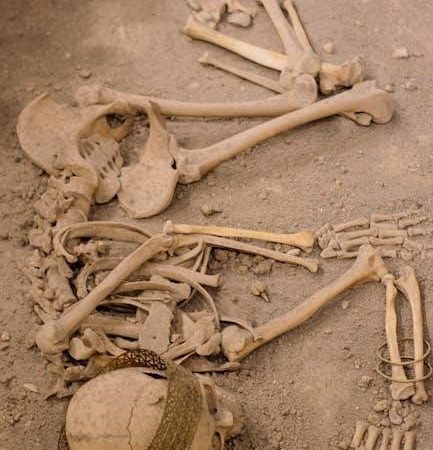
Human skeleton system PDFs offer detailed anatomical studies, showcasing the 206 bones and their functions—essential resources for students and medical professionals alike․
Overview of the Skeletal System
Human skeleton system PDFs are invaluable tools for understanding the body’s framework․ These resources detail the 206 bones, categorized into axial and appendicular components, providing crucial support and enabling movement․ Accessing comprehensive human skeleton system PDFs allows for detailed study of bone structures, joint types, and the overall organization of the skeletal system․
Such PDFs often include labeled diagrams, clinical correlations, and evolutionary comparisons, enhancing learning․ They are particularly useful for students in anatomy, physiology, and medical fields․ Reliable human skeleton system PDFs offer a visual and informative approach to mastering this complex biological system, aiding in diagnosis and treatment understanding․
The 206 Bones: A Fundamental Number
Human skeleton system PDFs consistently emphasize the significance of the 206-bone count in adult humans․ These resources meticulously detail each bone, categorizing them by region – skull, vertebral column, limbs, and girdles․ Accessing detailed human skeleton system PDFs allows for focused study of individual bone anatomy, including features like processes, foramina, and markings․
These PDFs often present the bones in a systematic manner, aiding memorization and comprehension․ Understanding this fundamental number is crucial for medical professionals and students․ High-quality human skeleton system PDFs provide clear illustrations and descriptions, solidifying knowledge of skeletal composition and its clinical relevance․
Axial vs․ Appendicular Skeleton
Human skeleton system PDFs clearly delineate the skeletal division into axial and appendicular components․ The axial skeleton, comprising 80 bones, forms the central core – skull, vertebral column, and rib cage – providing vital protection․ Appendicular skeleton PDFs detail the 126 bones of the limbs and their connecting girdles, enabling movement and flexibility․
Comprehensive human skeleton system PDFs often utilize comparative diagrams illustrating these distinctions․ Studying these resources enhances understanding of how each division contributes to overall body structure and function․ Detailed PDFs showcase the interconnectedness, emphasizing the axial skeleton’s support for the appendicular skeleton’s mobility․
The Axial Skeleton: Core Support
Human skeleton system PDFs illustrate the axial skeleton’s central role, detailing the skull, vertebral column, and rib cage’s protective and supportive functions․
Skull: Cranial and Facial Bones
Human skeleton system PDFs provide comprehensive views of the skull, differentiating between its cranial and facial components․ These resources meticulously detail the 22 cranial bones forming the cranium, protecting the brain, and the 14 facial bones constructing the face’s structure․
Detailed diagrams within these PDFs showcase the intricate sutures connecting these bones, alongside foramina—openings for nerves and blood vessels․ Students can utilize these visual aids to identify specific bones like the frontal, parietal, temporal, and occipital bones of the cranium․ Furthermore, PDFs highlight facial bones such as the maxilla, mandible, zygomatic, and nasal bones, crucial for understanding facial anatomy and potential fracture points․ Interactive 3D models, often linked within these PDFs, enhance spatial comprehension of the skull’s complex architecture․
Vertebral Column: Regions and Functions
Human skeleton system PDFs extensively illustrate the vertebral column’s structure and its vital functions․ These resources delineate the five regions: cervical, thoracic, lumbar, sacral, and coccygeal, each with unique characteristics․ Detailed diagrams showcase the curvature of the spine – lordosis and kyphosis – and their importance for balance and shock absorption․
PDFs often include labeled illustrations of typical vertebrae from each region, highlighting features like spinous and transverse processes․ They explain how the vertebral column protects the spinal cord and supports the body’s weight․ Interactive models within these PDFs allow users to explore the intervertebral discs and their role in flexibility and cushioning․ Understanding these regions is crucial, and PDFs provide the necessary visual and descriptive support․
Cervical Vertebrae: Anatomy and Alignment
Human skeleton system PDFs provide in-depth views of the cervical vertebrae, detailing their unique anatomy․ These resources emphasize the seven cervical vertebrae (C1-C7) and their specialized features, like the atlas (C1) and axis (C2), crucial for head movement․ PDFs illustrate the transverse foramina, allowing vertebral arteries to pass through, supplying the brain․
Detailed diagrams showcase the lordotic alignment of the cervical spine, explaining its importance for maintaining proper posture and balance․ Interactive 3D models within these PDFs allow for a comprehensive understanding of the cervical vertebrae’s articulation and range of motion․ PDFs also cover potential alignment issues and related pathologies․
Thoracic Vertebrae: Rib Cage Attachment
Human skeleton system PDFs meticulously illustrate the thoracic vertebrae (T1-T12) and their critical role in rib cage attachment․ These resources highlight the facets on the vertebral bodies and transverse processes where ribs articulate, forming a protective cage for vital organs․ Detailed diagrams showcase the varying rib attachment points along the thoracic spine․
PDFs often include labeled illustrations demonstrating how the rib cage contributes to respiration and spinal stability․ Interactive models allow users to visualize the movement of the ribs during breathing․ Furthermore, these resources explain the clinical significance of thoracic vertebral fractures and their impact on rib cage function, offering a comprehensive understanding․
Lumbar Vertebrae: Lower Back Support
Human skeleton system PDFs provide in-depth views of the lumbar vertebrae (L1-L5), emphasizing their robust structure designed for weight-bearing and lower back support․ These resources detail the large vertebral bodies and spinous processes characteristic of the lumbar region, crucial for maintaining upright posture․
PDFs often feature annotated diagrams illustrating the curvature of the lumbar spine and the surrounding musculature․ Interactive 3D models allow for a detailed examination of the intervertebral discs and their role in shock absorption․ Clinical notes within these PDFs frequently address common lumbar spine conditions like herniated discs and spinal stenosis․
Rib Cage: Protection of Vital Organs
Human skeleton system PDFs meticulously illustrate the rib cage’s protective function, showcasing how the ribs, sternum, and thoracic vertebrae safeguard vital organs like the heart and lungs․ Detailed anatomical charts within these resources highlight the true, false, and floating ribs, explaining their unique articulations․
PDFs often include cross-sectional diagrams demonstrating the rib cage’s relationship to the internal organs․ Interactive models allow users to visualize the expansion and contraction of the rib cage during respiration․ Clinical sections within these PDFs frequently address rib fractures and related injuries, offering valuable insights․

The Appendicular Skeleton: Movement and Flexibility
Human skeleton system PDFs detail limb bone structures, girdles, and joint mechanics, crucial for understanding human movement and flexibility through visual aids․
Upper Limbs: Bones of the Arms and Hands
Human skeleton system PDFs provide comprehensive illustrations and descriptions of the upper limb skeletal components․ These resources meticulously detail the humerus, radius, and ulna – the bones forming the arm’s framework․ Further exploration reveals the intricate structure of the carpus (wrist), comprised of eight small bones, enabling a wide range of motion․
Detailed PDFs also showcase the metacarpals, forming the palm, and the phalanges, constituting the fingers․ These documents often highlight key anatomical landmarks crucial for clinical applications․ Studying these PDFs allows for a deeper understanding of how these bones articulate to facilitate complex movements like grasping, lifting, and manipulating objects․ They are invaluable for students and professionals seeking a thorough grasp of upper limb anatomy․
Lower Limbs: Bones of the Legs and Feet
Human skeleton system PDFs offer detailed visual guides to the lower limb’s bony structure․ These resources clearly illustrate the femur, the longest and strongest bone in the human body, forming the thigh․ They also showcase the tibia and fibula, composing the lower leg, and their crucial role in weight-bearing and stability․
Furthermore, these PDFs meticulously depict the tarsals (ankle bones), metatarsals (foot bones), and phalanges (toe bones)․ They often include labeled diagrams highlighting important ligaments and joint surfaces․ Studying these resources enhances understanding of the lower limb’s biomechanics, essential for locomotion and supporting body weight․ These PDFs are invaluable for anatomical study and clinical practice․
Pectoral Girdle: Connecting Upper Limbs to the Axial Skeleton
Human skeleton system PDFs provide comprehensive views of the pectoral girdle, showcasing its vital connection between the upper limbs and the axial skeleton․ These resources detail the clavicle (collarbone) and scapula (shoulder blade), illustrating their unique shapes and articulating surfaces․
Detailed diagrams within these PDFs highlight the acromioclavicular and glenohumeral joints, crucial for upper limb mobility․ They often include annotations explaining the muscles attaching to these bones, influencing shoulder movement․ Studying these PDFs aids in understanding the girdle’s role in supporting the arm and enabling a wide range of motion, essential for anatomical comprehension․
Pelvic Girdle: Connecting Lower Limbs to the Axial Skeleton
Human skeleton system PDFs offer in-depth illustrations of the pelvic girdle, demonstrating its robust connection between the lower limbs and the axial skeleton․ These resources meticulously detail the hip bones – ilium, ischium, and pubis – and their fusion to form the pelvic bone․
PDFs often feature labeled diagrams showcasing the sacroiliac joints, where the pelvis connects to the sacrum, and the obturator foramen․ They clarify the girdle’s role in weight-bearing, locomotion, and protecting pelvic organs․ Studying these resources enhances understanding of the complex anatomy and biomechanics of this crucial skeletal structure․

Bone Development and Growth
Human skeleton system PDFs illustrate fetal cartilage models transforming into bone via osteoblasts and osteocytes, dependent on calcium and phosphorus for growth․
Fetal Cartilage Model
Human skeleton system PDFs extensively detail the initial stages of skeletal development, beginning with a foundational model constructed entirely of cartilage within the developing fetus․ These resources visually demonstrate how this cartilage serves as a blueprint for future bone formation․ The PDFs often include diagrams illustrating the progressive replacement of cartilage with bone tissue, a process orchestrated by specialized cells․
They highlight that this cartilaginous framework isn’t merely a temporary structure; it provides essential support and allows for growth before complete ossification occurs․ Detailed illustrations within these PDFs showcase the intricate shaping of future skeletal components during this crucial prenatal phase, emphasizing the importance of this initial cartilage model in establishing the overall skeletal structure․ Understanding this process is fundamental to comprehending skeletal anatomy and potential developmental variations․
Osteoblasts and Osteocytes: Bone Cell Roles
Human skeleton system PDFs thoroughly explain the dynamic roles of osteoblasts and osteocytes in bone development and maintenance․ These resources illustrate how osteoblasts, immature bone cells, actively synthesize and deposit new bone matrix, gradually replacing the initial cartilage model․ Detailed diagrams showcase osteoblasts’ crucial function in building bone tissue during growth and repair․
Furthermore, PDFs clarify that once osteoblasts become embedded within the bone matrix, they mature into osteocytes․ These osteocytes then maintain the bone, ensuring its continued health and structural integrity․ The resources emphasize that osteocytes are vital for sensing mechanical stress and regulating bone remodeling, highlighting their ongoing contribution to skeletal function throughout life․
Mineral Dependence: Calcium and Phosphorus
Human skeleton system PDFs consistently emphasize the critical dependence of bone formation on adequate mineral supply, particularly calcium (Ca) and phosphorus (P)․ These resources detail how these minerals are integral components of hydroxyapatite, the primary mineral salt that provides bone its hardness and rigidity․ Visual aids within the PDFs illustrate the chemical composition of bone and the role of these minerals․
Furthermore, the documents explain that insufficient intake of calcium or phosphorus can lead to weakened bones and increased susceptibility to fractures․ They often include dietary recommendations and discuss conditions like osteoporosis, linked to mineral deficiencies, providing a comprehensive understanding of this vital relationship․
Long Bones: Majority Bone Type
Human skeleton system PDFs frequently highlight that long bones constitute the majority of the skeletal structure․ These resources detail their characteristic features – a shaft (diaphysis) and two ends (epiphyses) – and their primary function in facilitating movement․ Diagrams within these PDFs clearly illustrate examples like the femur, tibia, and humerus, showcasing their elongated shape․
The documents explain how long bones contain bone marrow, crucial for blood cell production, and are sites for muscle attachment․ They also cover growth plate activity during development, demonstrating how these bones lengthen․ Studying these PDFs provides a thorough understanding of long bone anatomy and physiology․

Bone Structure and Composition
Human skeleton system PDFs illustrate that bones comprise compact and spongy tissue, alongside bone marrow—vital for understanding skeletal strength and functionality․
Compact Bone: Density and Strength
Human skeleton system PDFs meticulously detail compact bone, the dense outer layer providing significant strength and protection․ These resources visually demonstrate its organized structure – Haversian systems, or osteons – containing central canals with blood vessels and nerves․
PDFs highlight how this tightly packed arrangement of collagen fibers and mineral salts (calcium and phosphorus) contributes to bone’s remarkable resistance to stress․ They often include microscopic images showcasing lamellae, concentric rings around the central canals․ Understanding compact bone’s composition, as presented in these PDFs, is crucial for grasping how the skeleton withstands daily forces and protects vital organs․ Detailed diagrams illustrate the interplay between osteocytes within lacunae and canaliculi, facilitating nutrient exchange․
Spongy Bone: Lightweight Support
Human skeleton system PDFs extensively cover spongy bone, also known as cancellous bone, found primarily at the ends of long bones and within vertebrae․ These resources illustrate its porous, network-like structure, composed of trabeculae – small, irregular plates of bone․
PDFs emphasize that this architecture, while less dense than compact bone, provides crucial lightweight support and creates space for bone marrow․ Detailed diagrams showcase how trabeculae align along lines of stress, maximizing strength with minimal weight․ Understanding the function of spongy bone, as detailed in these PDFs, is vital for comprehending shock absorption and efficient movement․ They often include microscopic views highlighting the interconnected network and bone marrow cavities․
Bone Marrow: Red and Yellow Types
Human skeleton system PDFs dedicate significant sections to bone marrow, detailing its two primary types: red and yellow․ These resources illustrate that red marrow, found mainly in flat bones and epiphyses of long bones, is responsible for hematopoiesis – the production of blood cells․
PDFs clearly explain the transition from predominantly red marrow in youth to an increase in yellow marrow with age․ Yellow marrow, largely composed of fat, serves as an energy reserve․ Detailed diagrams within these PDFs showcase the microscopic structure of both types, highlighting the differences in cellular composition․ Understanding the roles of each marrow type, as presented in these PDFs, is crucial for grasping overall skeletal function and health․
Joints: Where Bones Meet
Human skeleton system PDFs comprehensively cover joint types—fibrous, cartilaginous, and synovial—explaining their structures and functions with detailed illustrations and explanations․
Types of Joints: Fibrous, Cartilaginous, Synovial
Human skeleton system PDFs meticulously detail the three major joint classifications․ Fibrous joints, like sutures in the skull, offer limited movement and strong connections․ Cartilaginous joints, such as those in the vertebral column, permit slight movement and provide cushioning․
Synovial joints, the most common type, enable a wide range of motion—found in limbs․ These PDF resources illustrate synovial joint structures: articular cartilage, joint capsules, and synovial fluid․ They explain how these components facilitate smooth movement while minimizing friction․ Detailed diagrams showcase examples like hinge, ball-and-socket, and pivot joints, clarifying their unique mechanics and range of motion․ Understanding these classifications is crucial for comprehending skeletal function and potential pathologies․
Synovial Joints: Structure and Function

Human skeleton system PDFs extensively cover synovial joints, highlighting their complex structure․ These joints feature articular cartilage reducing friction, a joint capsule enclosing the joint, and synovial fluid providing lubrication and nourishment․ Ligaments reinforce the capsule, enhancing stability․
PDF resources detail how these components work in concert to enable movement․ Muscles attach via tendons, generating force․ The PDFs illustrate the role of bursae—fluid-filled sacs—minimizing friction․ Understanding synovial joint function is vital for diagnosing conditions like arthritis․ Diagrams clearly depict the interplay of these structures, aiding comprehension of joint mechanics and potential injury points․

Skeletal System in Evolutionary Anatomy
Human skeleton system PDFs reveal comparative anatomy, tracing skeletal changes across species—illuminating our evolutionary path and mammalian skeletal distinctions․
Human vs․ Mammalian Skeletons
Human skeleton system PDFs are invaluable when comparing our skeletal structure to that of other mammals․ These resources highlight key differences, such as bipedalism’s impact on the human pelvis and vertebral column, creating unique angles and curvatures not typically found in quadrupedal mammals․
Detailed illustrations within these PDFs demonstrate variations in limb proportions, skull morphology, and the overall bone density between species․ For instance, Merrilees and Porter (1979), referenced in evolutionary anatomy studies, provide a guide to identifying Australian mammal skeletons․ Analyzing these PDFs reveals how skeletal adaptations reflect lifestyle and environmental pressures, showcasing the remarkable diversity within the mammalian class and pinpointing the specific evolutionary trajectory of Homo sapiens․
Comparative Skeletal Anatomy
Human skeleton system PDFs facilitate detailed comparative analyses, revealing subtle yet significant anatomical distinctions across species․ These resources allow for side-by-side examination of bone structures, highlighting variations in size, shape, and articulation points․ Studying these PDFs demonstrates how skeletal features correlate with locomotion, diet, and environmental adaptation․
For example, PDFs often include detailed illustrations comparing human hand bones to those of primates, showcasing adaptations for grasping versus manipulation․ Examining pelvic girdle structures in different mammals reveals how skeletal morphology supports varying modes of reproduction and movement․ These comparative studies, readily available in PDF format, are crucial for understanding evolutionary relationships and the functional significance of skeletal anatomy․

Common Skeletal System Conditions
Human skeleton system PDFs illustrate conditions like osteoporosis and arthritis, detailing bone density loss and joint inflammation through diagnostic imaging and anatomical explanations․
Osteoporosis: Bone Density Loss
Human skeleton system PDFs are invaluable resources for understanding osteoporosis, a condition characterized by diminished bone density and increased fracture risk․ These documents often feature comparative imaging – X-rays and scans – demonstrating healthy bone structure versus osteoporotic bone, visually highlighting the loss of trabecular network․
Detailed anatomical illustrations within these PDFs pinpoint areas most susceptible to fractures, such as the hip, spine, and wrist․ Furthermore, they explain the cellular mechanisms involved, detailing how imbalances between osteoblast and osteoclast activity contribute to bone weakening․ Many PDFs also cover risk factors, diagnostic procedures, and preventative measures, including calcium and vitamin D supplementation, making them comprehensive guides for both patients and healthcare providers seeking in-depth knowledge of this prevalent skeletal condition․
Arthritis: Joint Inflammation
Human skeleton system PDFs provide crucial insights into arthritis, a condition involving joint inflammation and pain․ These resources often showcase detailed diagrams illustrating the various types of arthritis – osteoarthritis, rheumatoid arthritis, and gout – and their specific impacts on joint structures like cartilage and synovial membranes․
PDFs frequently include radiographic images demonstrating joint space narrowing, bone spurs, and erosions characteristic of arthritic changes․ They explain the underlying pathophysiology, detailing the immune responses and cartilage degradation involved․ Many also cover treatment options, from pharmacological interventions to physical therapy, offering a comprehensive understanding of managing this debilitating condition․ These PDFs are essential for students and clinicians alike, aiding in accurate diagnosis and effective patient care․

Resources for Further Study (PDF Focus)
Human skeleton system PDFs are readily available online, offering in-depth anatomical charts, clinical case studies, and comprehensive guides for detailed learning․
Finding Reliable Human Skeleton System PDFs
Locating trustworthy human skeleton system PDFs requires careful source evaluation․ University websites and medical institutions frequently offer high-quality, peer-reviewed anatomical resources in PDF format․ Search for publications from established anatomy departments or medical schools; these are generally reliable․
Avoid websites with unclear authorship or excessive advertising․ Reputable publishers of medical textbooks often provide supplementary PDF materials for instructors and students․ Online libraries and digital archives, like PubMed Central, can also yield valuable PDFs․
Always verify the PDF’s date to ensure the information is current, as anatomical knowledge evolves․ Cross-reference information with multiple sources to confirm accuracy and completeness․ Prioritize PDFs that cite credible sources and are authored by qualified professionals․
Utilizing PDF Resources for Anatomy Learning
Human skeleton system PDFs are invaluable for detailed study․ Utilize interactive features like hyperlinks within the PDF to navigate between sections and explore related anatomical structures․ Zoom functions allow close examination of bone details and joint articulations․
Annotate PDFs with notes, labels, and diagrams to reinforce learning․ Combine PDF study with 3D anatomical models for a comprehensive understanding․ Regularly review the material and test your knowledge with quizzes or self-assessment questions․
Focus on understanding the functional relationships between bones and how they contribute to movement and support․ Remember to cross-reference PDF information with other learning resources for a holistic perspective․