Image Guided Superficial Radiation Therapy: A Comprehensive Overview
Image-guided superficial radiation therapy (IGSRT) represents a significant advancement, offering precise treatment for skin cancers and enhancing
palliative care through real-time imaging and optimized dose delivery, minimizing risks.
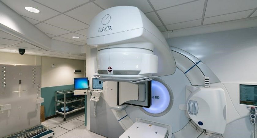
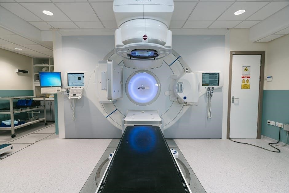
Image-Guided Superficial Radiation Therapy (IGSRT) is an evolving technique in radiation oncology, specifically designed for treating superficial tumors, primarily skin cancers. It combines the principles of traditional superficial radiation therapy with advanced imaging technologies to dramatically improve treatment precision and accuracy. Unlike conventional methods, IGSRT utilizes real-time imaging – such as orthogonal imaging, cone-beam computed tomography (CBCT), or surface-guided radiation therapy (SGRT) – to verify patient positioning and tumor localization before and sometimes during each treatment session.
This capability is crucial for minimizing dose to surrounding healthy tissues, a key advantage over older techniques. IGSRT is increasingly employed for basal cell carcinoma and squamous cell carcinoma, offering a non-invasive alternative to surgery, and also finds application in palliative care scenarios. The integration of imaging allows for adaptive treatment planning, tailoring the radiation dose to the individual patient’s anatomy and tumor characteristics, ultimately leading to improved clinical outcomes and reduced side effects;
Historical Development of Superficial Radiation Therapy
Superficial radiation therapy (SRT) has a long history, originating in the early 20th century with the discovery of X-rays. Initially, SRT utilized techniques with limited precision, often relying on manual setup and palpation for tumor localization. Early applications focused on treating skin cancers, but dose control and minimizing collateral damage were significant challenges. Over decades, advancements in radiation delivery systems, like orthovoltage X-ray machines, improved dose distribution.
However, a pivotal shift occurred with the introduction of image guidance. The integration of imaging modalities – initially radiography, and later CBCT and SGRT – revolutionized SRT, transforming it into Image-Guided SRT (IGSRT). This evolution enabled real-time verification of patient positioning and tumor targeting, dramatically enhancing accuracy and reducing treatment-related toxicity. Ongoing research, including investigations into radiotherapy deintensification, continues to refine IGSRT, building upon its historical foundations.
The Role of Imaging in Radiation Therapy Precision
Imaging is fundamental to the precision of modern radiation therapy, particularly in Image-Guided Superficial Radiation Therapy (IGSRT). Historically, treatment relied on physical landmarks, prone to inaccuracies due to patient movement or anatomical variations. The advent of imaging technologies – including orthogonal imaging, Cone-Beam Computed Tomography (CBCT), and Surface Guided Radiation Therapy (SGRT) – allows for real-time visualization of the tumor and surrounding tissues.
This capability enables precise target delineation and accurate dose delivery, minimizing exposure to healthy structures. Imaging confirms correct patient positioning before and during each treatment fraction, correcting for any deviations. Furthermore, image guidance supports treatment adaptation based on observed tumor response. By integrating imaging, IGSRT significantly improves treatment efficacy and reduces the risk of adverse effects, enhancing overall patient outcomes and safety.
Principles of IGSRT
IGSRT’s core principles involve utilizing real-time imaging – orthogonal, CBCT, or SGRT – to precisely target superficial tumors, ensuring accurate dose delivery and minimizing collateral damage.
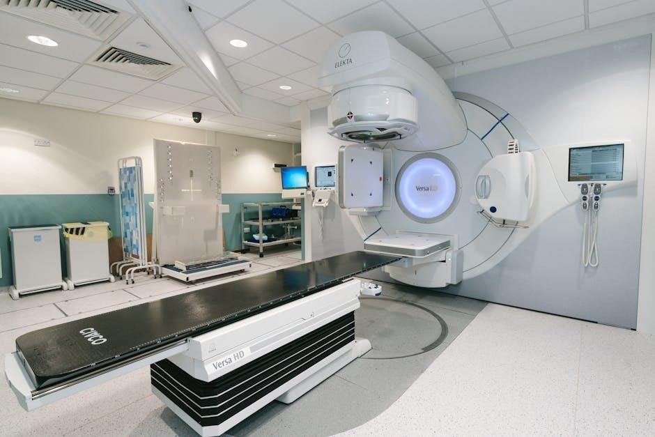
Real-Time Image Guidance Techniques
Real-time image guidance is fundamental to IGSRT, enabling precise tumor localization and treatment verification before and during each radiation fraction. This dynamic approach contrasts with traditional methods, significantly improving accuracy. Techniques include orthogonal imaging, providing two-dimensional views for setup confirmation, and cone-beam computed tomography (CBCT), offering three-dimensional volumetric imaging for detailed anatomical assessment.
Surface Guided Radiation Therapy (SGRT) represents another crucial technique, utilizing cameras to capture the patient’s surface topography and compare it to a reference scan, allowing for real-time adjustments to patient positioning. These techniques collectively address inter-fractional and intra-fractional motion, ensuring the radiation beam consistently targets the intended volume, while sparing surrounding healthy tissues. The integration of these technologies minimizes setup errors and enhances treatment precision, ultimately leading to improved clinical outcomes.
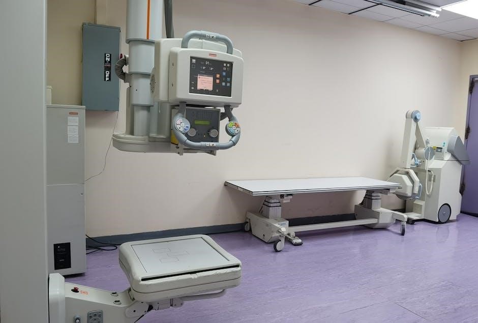
Orthogonal Imaging and Cone-Beam Computed Tomography (CBCT)
Orthogonal imaging, employing paired radiographic images, serves as a rapid and straightforward method for daily patient setup verification in IGSRT. It confirms bony anatomy alignment, ensuring consistent beam delivery. However, its two-dimensional nature offers limited soft tissue visualization. Cone-Beam Computed Tomography (CBCT) overcomes this limitation, providing a three-dimensional volumetric image of the treatment area.
CBCT allows for detailed assessment of tumor and surrounding tissue positions, crucial for accurate dose calculation and delivery. It’s particularly valuable for detecting subtle anatomical changes between fractions. While CBCT involves a higher radiation dose than orthogonal imaging, its enhanced precision justifies its use in complex cases. Both modalities contribute to minimizing setup errors and maximizing target coverage, ultimately improving treatment efficacy and reducing side effects in IGSRT.
Surface Guided Radiation Therapy (SGRT)
Surface Guided Radiation Therapy (SGRT) utilizes cameras and infrared light to capture the patient’s surface anatomy, creating a 3D representation that’s matched to the original treatment plan. This real-time monitoring allows for precise patient positioning before and during each radiation fraction, accounting for daily variations caused by breathing, weight fluctuations, or patient movement.
SGRT is particularly beneficial for superficial targets where soft tissue shifts significantly impact accuracy. It minimizes the need for implanted markers, enhancing patient comfort. By continuously verifying the patient’s position, SGRT ensures the radiation beam consistently targets the intended area, reducing dose to healthy tissues. This technology complements CBCT and orthogonal imaging, providing a comprehensive image guidance solution for optimized IGSRT delivery.

Clinical Applications of IGSRT
IGSRT excels in treating basal and squamous cell carcinomas, offering effective palliative care for skin cancers, and improving outcomes with precise, targeted radiation delivery.
Treatment of Basal Cell Carcinoma
Image-Guided Superficial Radiation Therapy (IGSRT) provides a non-invasive alternative to surgical excision for basal cell carcinoma (BCC), particularly beneficial for patients unsuitable for surgery or desiring cosmetic preservation. IGSRT’s precision minimizes damage to surrounding healthy tissues, crucial for delicate areas like the face. Real-time imaging ensures accurate targeting, adapting to anatomical variations and potential movement during treatment.
The technique is especially valuable for treating multiple BCCs simultaneously, streamlining the treatment process. IGSRT’s shallow depth of penetration is ideal for BCC, a cancer typically confined to the epidermis and superficial dermis. Treatment plans are carefully optimized based on tumor size, location, and patient-specific factors, maximizing efficacy while minimizing side effects. Ongoing monitoring during treatment allows for adjustments, further enhancing precision and control.
Management of Squamous Cell Carcinoma
Image-Guided Superficial Radiation Therapy (IGSRT) offers a compelling treatment option for Squamous Cell Carcinoma (SCC), particularly for lesions in challenging anatomical locations or for patients with comorbidities. IGSRT’s precision is vital, as SCC has a greater potential for local recurrence and, in rare cases, metastasis compared to basal cell carcinoma. Real-time imaging guides accurate dose delivery, accounting for tumor depth and surrounding critical structures.
IGSRT allows for tailored treatment plans, adjusting for tumor size, location, and histological grade. The technique is effective for both primary SCCs and recurrent lesions following surgical removal. Careful patient selection and meticulous treatment planning are essential to maximize efficacy and minimize the risk of complications. IGSRT provides a non-invasive alternative, preserving function and cosmesis.
Palliative Care Applications in Skin Cancer
Image-Guided Superficial Radiation Therapy (IGSRT) plays a crucial role in palliative care for advanced or metastatic skin cancers, significantly improving quality of life for patients. IGSRT effectively manages symptomatic lesions, such as ulcerated tumors causing pain, bleeding, or obstruction, offering rapid symptom relief with minimal invasiveness. Precise targeting minimizes dose to healthy tissues, reducing side effects crucial for patients with compromised health.
The technique is particularly valuable for patients ineligible for or declining curative treatments. Short treatment courses are often sufficient to achieve symptom control, enhancing comfort and functionality. IGSRT allows for compassionate care, addressing patient needs and improving overall well-being during end-of-life stages. It represents a valuable tool in holistic skin cancer management.
Treatment Planning and Delivery
IGSRT utilizes meticulous dose calculations, patient immobilization, and optimized fractionation strategies to ensure accurate and effective radiation delivery for targeted skin cancers.
Dose Calculation and Optimization
Precise dose calculation is paramount in IGSRT, leveraging advanced algorithms to accurately model radiation distribution within the superficial tissues. Optimization focuses on maximizing the dose to the targeted lesion while simultaneously minimizing exposure to surrounding healthy skin and critical structures. This involves careful consideration of beam angles, energy levels, and treatment parameters.
The goal is to achieve a therapeutic ratio that effectively eradicates cancerous cells with minimal collateral damage. IGSRT’s image guidance allows for real-time adjustments to the treatment plan, ensuring conformity to the tumor volume even with anatomical changes during the course of therapy. Sophisticated planning systems aid clinicians in visualizing dose distributions and predicting potential side effects, leading to highly individualized and optimized treatment strategies.
Patient Positioning and Immobilization
Accurate and reproducible patient positioning is crucial for IGSRT’s effectiveness. Before each treatment fraction, patients are positioned using the image guidance system, ensuring alignment with the original treatment plan. Immobilization devices, such as custom-fitted masks or cushions, may be employed to minimize movement during treatment delivery.
These devices help maintain consistent geometry, compensating for patient motion and anatomical variations. Real-time surface monitoring, a key component of SGRT, further verifies patient position before and during radiation delivery. This meticulous approach ensures that the radiation beam precisely targets the intended tumor volume, maximizing therapeutic benefit and minimizing dose to healthy tissues. Proper positioning reduces setup errors and enhances treatment precision.

Treatment Fractionation Strategies
Treatment fractionation in IGSRT involves dividing the total radiation dose into smaller, daily fractions delivered over several weeks. This approach balances tumor control probability with minimizing acute and late toxicities. Common fractionation schemes for superficial skin cancers include delivering 50-60 Gy in 10-20 fractions, though protocols are tailored based on tumor characteristics and location.
IGSRT allows for dose escalation to the tumor while sparing surrounding healthy tissues, potentially enabling hypofractionation – delivering fewer, larger fractions. Research explores radiotherapy deintensification, reducing doses for low-risk patients. Careful consideration of fractionation is vital for optimizing treatment outcomes and patient quality of life, guided by imaging feedback.
Benefits and Advantages of IGSRT
IGSRT delivers superior accuracy, improved target delineation, and reduced doses to healthy tissues, resulting in enhanced patient comfort and significantly shortened treatment times.
Improved Accuracy and Target Delineation
Image guidance fundamentally elevates the precision of superficial radiation therapy, enabling clinicians to accurately define and target tumor margins with unprecedented detail. Techniques like orthogonal imaging and cone-beam computed tomography (CBCT), alongside surface guided radiation therapy (SGRT), provide real-time visualization of the treatment area.
This capability is crucial for skin cancers, where precise targeting minimizes damage to surrounding healthy tissue. Accurate delineation ensures the radiation dose is concentrated on the malignancy, maximizing therapeutic effect while sparing normal structures. Furthermore, image guidance accounts for patient movement during treatment, dynamically adjusting the beam to maintain accuracy throughout each session. This leads to more consistent and reliable dose delivery, ultimately improving treatment outcomes and reducing the potential for side effects.
Reduced Dose to Healthy Tissue
A core benefit of image-guided superficial radiation therapy (IGSRT) lies in its ability to significantly reduce radiation exposure to surrounding healthy tissues. By utilizing real-time imaging – including orthogonal imaging, CBCT, and SGRT – clinicians can precisely conform the radiation beam to the tumor volume, minimizing scatter and penumbra.
This targeted approach is particularly important in superficial treatments, where tumors are close to the skin surface and critical structures. Accurate patient positioning and immobilization, coupled with continuous image verification, further contribute to dose sparing. Consequently, IGSRT reduces the incidence and severity of acute skin reactions and long-term skin changes, enhancing patient comfort and quality of life during and after treatment. This precision is vital for optimizing therapeutic ratios.
Enhanced Patient Comfort and Reduced Treatment Time
Image guidance streamlines the superficial radiation therapy process, leading to notable improvements in patient comfort and a reduction in overall treatment duration. Precise targeting minimizes the need for extensive treatment fields, decreasing the volume of normal tissue exposed to radiation. This focused approach translates to fewer treatment fractions in some cases, lessening the burden on patients.
Furthermore, real-time imaging allows for quick adjustments to patient positioning, ensuring accuracy without prolonged setup times. The non-invasive nature of SGRT, in particular, contributes to a more comfortable experience. Reduced treatment times also improve workflow efficiency within radiation oncology departments, allowing for greater patient throughput and access to care.

Potential Risks and Side Effects
IGSRT may cause acute skin reactions and, less commonly, long-term skin changes; careful risk assessment and mitigation strategies are crucial for patient safety.
Acute Skin Reactions
Acute skin reactions are the most common side effect of image-guided superficial radiation therapy (IGSRT), typically manifesting as redness, dryness, itching, and peeling within the treated area. These reactions are generally similar to a mild sunburn and usually appear during or shortly after the completion of treatment. The severity can vary depending on the dose, treatment duration, and individual patient factors.
Effective management of these reactions often involves gentle skin care, including the use of bland, fragrance-free moisturizers and avoidance of harsh soaps or scrubbing. Topical corticosteroids may be prescribed to alleviate inflammation and itching. Patients are generally advised to avoid direct sun exposure to the treated area and to wear protective clothing. Most acute skin reactions resolve within a few weeks following the completion of IGSRT, and rarely require interruption of treatment.
Long-Term Skin Changes
Long-term skin changes following image-guided superficial radiation therapy (IGSRT) are less common than acute reactions, but can occur in some patients. These may include persistent dryness, altered skin pigmentation (either darkening or lightening), thinning of the skin, or telangiectasias (small, visible blood vessels). In rare cases, radiation-induced fibrosis or ulceration can develop, particularly with higher doses or in patients with pre-existing skin conditions.
The risk of long-term changes is minimized through careful treatment planning and dose optimization, ensuring precise targeting of the tumor while sparing surrounding healthy tissue. Regular follow-up appointments are crucial for monitoring skin health and addressing any concerns. Proactive skin care, including consistent moisturizing and sun protection, can help mitigate potential long-term effects and maintain skin integrity.
Risk Assessment and Mitigation Strategies
Comprehensive risk assessment is paramount before initiating image-guided superficial radiation therapy (IGSRT). This includes evaluating patient history, skin condition, and tumor characteristics to predict potential adverse effects. Mitigation strategies begin with meticulous treatment planning, utilizing precise imaging to minimize dose to healthy tissues. Careful patient positioning and immobilization are also crucial for accuracy.
Proactive management of acute skin reactions, such as dryness and redness, with emollients and wound care, reduces the likelihood of complications. Educating patients about potential side effects and proper skin care is essential. Regular follow-up appointments allow for early detection and intervention if long-term changes develop, ensuring optimal outcomes and patient well-being.
Emerging Technologies and Future Directions
Artificial intelligence (AI) integration, alongside intraoperative radiation therapy (IORT), promises radiotherapy deintensification, improving precision and outcomes in image-guided superficial radiation therapy.
Artificial Intelligence (AI) in IGSRT
Artificial intelligence (AI) is poised to revolutionize image-guided superficial radiation therapy (IGSRT) across multiple facets of the treatment pathway. AI algorithms can automate target volume delineation, significantly reducing inter-observer variability and improving accuracy in defining the treatment area. Furthermore, AI-powered image analysis can enhance the identification of subtle tumor margins, particularly crucial in skin cancer management.
Predictive modeling, utilizing machine learning, can personalize treatment plans by forecasting potential skin reactions and optimizing dose distributions based on individual patient characteristics. This leads to reduced toxicity and improved cosmetic outcomes. AI also facilitates real-time adaptive planning, adjusting treatment parameters during delivery based on observed anatomical changes.
Beyond treatment planning, AI can streamline quality assurance processes, automatically detecting inconsistencies and ensuring adherence to established protocols. The integration of AI promises to enhance efficiency, precision, and personalization within IGSRT, ultimately leading to superior patient care.
Integration with Intraoperative Radiation Therapy (IORT)
Combining image-guided superficial radiation therapy (IGSRT) with intraoperative radiation therapy (IORT) presents a compelling strategy for enhanced cancer control, particularly in cases requiring immediate post-surgical treatment. IORT delivers a concentrated radiation dose directly to the tumor bed after surgical resection, minimizing exposure to surrounding healthy tissues. Integrating IGSRT pre-operatively allows for precise tumor localization and margin assessment, informing the IORT delivery plan.
This synergistic approach is particularly valuable in high-risk skin cancers where complete surgical excision is challenging. Real-time imaging during IORT, guided by pre-operative IGSRT data, ensures accurate targeting and dose conformity. Research indicates potential benefits in radiotherapy deintensification, reducing overall treatment duration and toxicity for low-risk patients.
Furthermore, the combined modality offers a potentially curative option for patients unsuitable for traditional external beam radiation therapy. The future of cancer treatment lies in such integrated approaches, maximizing efficacy while minimizing side effects;
Research on Radiotherapy Deintensification
Radiotherapy deintensification, leveraging the precision of image-guided superficial radiation therapy (IGSRT), is a growing area of research focused on minimizing treatment toxicity without compromising cancer control. Traditional radiation often delivers doses exceeding what’s biologically necessary, leading to unwanted side effects. IGSRT’s accurate targeting allows for dose reduction, particularly in favorable cases.
Studies, like the RTOG-0630 trial examining image-guided radiation for low-risk patients, demonstrate the feasibility of reducing target volumes and overall doses. This approach is especially relevant for superficial skin cancers where precise delineation is achievable. Combining IGSRT with intraoperative radiation therapy (IORT) further facilitates deintensification by delivering a concentrated boost directly to the tumor bed.
Ongoing research explores biomarkers to identify patients most likely to benefit from reduced doses, personalizing treatment and maximizing therapeutic ratios.